The Center for Translational Pathology at New York-Presbyterian/Weill Cornell Medicine provides histology, immunohistochemistry, tissue microarray construction and use, molecular pathology, multiplex immunofluorescence, whole slide scanning services and computational pathology to support the translational research of the department's investigators.
Histology
Histology related services include fixation, processing, embedding, and sectioning of human and animal tissues. We provide formalin fixed paraffin embedded (FFPE) and frozen embedding in OCT services for routine histology. Routine H&E staining and special stains are also performed in our Pathology Core Laboratory. We can advise researchers on tissue preparation, preservation and handling to ensure high quality results.
Automated Vacuum Tissue Processor
The Leica ASP6025 is a modular tissue processor for the following laboratory applications: fixation, dehydration, infiltration with intermedium, and the paraffin infiltration of histological tissue specimens.
Immunohistochemistry
Services include automated immunohistochemical labeling of established antibodies, and optimization of new antibodies with a variety of detection systems is performed. The detection system most frequently used is the polymer-HRP system using DAB with Hematoxylin counterstain.
Bond III & Bond Rx Autostainer
The Leica Autostainer is capable of running multiple IHC protocols simultaneously with, or without routine de-paraffination and pretreatment. Protocols are 100% customizable and multiple chromagens can be used side by side. The automation of the system provides highly specific and reproducible results. Recent upgrades to this equipment now allow for double staining and triple staining within the same protocol to examine co-expression. Service can be requested through CTP website.
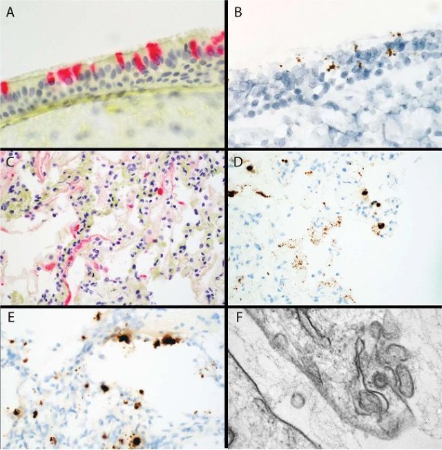
Immunohistochemistry and RNAish for viral spike protein in tracheal epithelium. Modern Pathology volume 33, pages 2156–2168(2020)
Micro Tissue Array
The tissue microarray instruments allow generation of multiple specimen slides that contain hundreds of individual tissues. Instead of incubating and analyzing samples one slide at a time, tissue microarrays allow you to examine hundreds of samples within a single slide. An immunohistochemical analysis from 400 different tissues can be completed in a day. All the histochemical and molecular detection techniques that can be used with regular sections can also be used with tissue microarrays with increased speed, throughput, standardization and conservation of valuable tissue.
RNA in situ hybridization
- RNAScope: The RNAScope assay represents a revolutionary advance in in situ target mRNA and ncRNA targets of >300bp. CTP is offering chromogenic (DAB) detection on the automated Leica Bond system.
- BaseScope: The BaseScopeTM Assay enables applications such as the detection of exon junctions/splice variants, short/highly homologous RNA sequences (50-300 bases), and point mutations at single cell sensitivity. CTP is offering detection of human BRAF (V600E), EGFR (L858R & T790M), KRAS (G12A, G12C, G12D, G12S, G12V) and PIK3CA (H1047R) mutations on FFPE tissue sections by using Leica Bond automated stainer.
Brightfield Slide Scanning
The Aperio GT450 and AT Turbo are capable to scan up to 400 slides within one load and convert glass slides to digital slide files, which are then stored in a database for access by our Pathology researchers. With dedicated workflows for research and biopharma coupled with an intuitive interface, Aperio eSlide Manager is the ideal solution to meet the diverse needs of both entry-level and enterprise digital pathology users.
Quantitative Image Analysis
A workstation with inform tissue analysis and HALO imaging analysis software is available for users to request independent use. InForm is a patented automated image analysis software package for accurately visualizing and quantifying biomarkers in tissue sections. HALO software, including six modules (TMA, Classifier, Cytonuclear IHC, Membrane IHC, ISH & Highplex FL), is available for image analysis. Service can be requested through CTP website.
Vectra Polaris and CODEX System
The Vectra Polaris Automated Quantitative Pathology Imaging System is a new class of tissue imager which provides unparalleled speed, performance, and versatility for visualizing, analyzing, quantifying, and phenotyping immune cells in situ in Formalin Fixed Paraffin Embedded (FFPE) tissue sections and TMAs to advance disease research. Vectra Polaris goes beyond basic functions, like whole-slide, brightfield (BF), and fluorescence (FL) imaging. It integrates the power of multispectral imaging in a simplified workflow.
GeoMx Digital Spatial Profiler
GeoMx Digital Spatial Profiler is a novel platform developed by NanoString. Digital Spatial Profiling is based on the nCounter® barcoding technology that enables spatially resolved, digital characterization of proteins or mRNA in a highly multiplexed (up to 1,000-plex) assay. The assay relies upon antibody or RNA probes coupled to photocleavable oligonucleotide tags. After hybridization of probes to slide-mounted formalin fixed paraffin-embedded (FFPE) tissue sections, the oligonucleotide tags are released from discrete regions of the tissue via UV exposure. Released tags are quantitated in a standard nCounter assay, and counts are mapped back to tissue location, yielding a spatially-resolved digital profile of analyte abundance.
Flow Cytometer LSR II & FACSAria Fusion Cell Sorter
Pathology BD LSR II is a powerful air-cooled three-laser benchtop flow cytometer with the ability to acquire parameters for ten different colors. The BD FACSAria™ Fusion is a flow cytometer that provides cell sorting. The instrument performs the tasks required for flow cytometry using BD FACSDiva™ software. The software provides the setup, acquirement, and analysis tasks with only minimal intervention required from the operator. This software is compatible with common BD cell sorters and analyzers. This enables researchers to utilize other systems for specific tasks without having to worry about incompatibility.
Computational Pathology
(Coming Soon)